- What is an abdominal ultrasound?
- Why would I need an abdominal ultrasound?
- How do I prepare for an abdominal ultrasound?
- Does an abdominal ultrasound hurt?
- Is an abdominal ultrasound better than an X-ray?
An abdominal ultrasound is a non-invasive diagnostic tool doctors use to visualize inside your body. As a screening tool, many doctors use ultrasounds for a wide range of purposes, from imaging pregnancies to identifying vascular conditions.
Since our vessels are composed of soft tissues, ultrasounds are one of the preferred diagnostic tools used by vascular surgeons, who interpret the imaging results to identify issues affecting your veins and arteries.
While receiving an ultrasound is a common and standard diagnostic process, patients who have never received one before often have questions about how it works.
Adam B. Levitt, M.D., R.V.T., F.A.C.S., board-certified vascular surgeon at Vascular Specialists of Central Florida says, “Patients commonly ask us about the ultrasound process, what it involves, and what they should expect.”
In this article, we’ll help you understand what you can expect from an abdominal ultrasound, how vascular surgeons use ultrasound technology, and why you can benefit from having one.
What Is an Abdominal Ultrasound?
 An abdominal ultrasound is a diagnostic test that uses high-frequency sound waves to create images of your organs and other structures inside the abdomen. As the sound waves bounce off the tissue in your body, the sensors on the ultrasound machine interpret this data, generating an image based on the composition and intensity of the feedback.
Thanks to the use of sound waves, the procedure is non-invasive and can be performed while the patient is seated comfortably in a chair.
For patients of Dr. Levitt, “You’ll be greeted professionally and then seen by one of our vascular technologists. They’ll bring you into the room and put a probe on your stomach to look at your arteries or veins.”
That small handheld probe is called a transducer. The transducer emits painless sound waves into the belly. These soundwaves bounce back, and a computer processes the echoes to create images of your internal organs, highlighting specific areas of the body:
- Bladder
- Gallbladder
- Kidneys
- Liver
- Pancreas
- Spleen
The abdominal ultrasound also captures information from the surrounding structures, including the arteries and veins that feed blood to and from these vital organs. The images doctors capture provide valuable information about these structures’ shape, size, and texture. Vascular surgeons like Dr. Levitt can look for abnormalities restricting blood flow, such as blockages or changes in the size of the vessels such as aneurysms.
Why Would I Need an Abdominal Ultrasound?
 The abdominal ultrasound is a standard tool used by many types of doctors, including vascular surgeons like Dr. Levitt, who says, “we might use an ultrasound machine to look for an aortic aneurysm, first looking for renal artery stenosis, meaning a narrowing to your kidney arteries.”
An aortic aneurysm is a serious condition where the walls of the aorta weaken or bulge. The aorta is the largest artery in the body. It carries oxygen-rich blood from the heart to the rest of our organs.
Renal artery stenosis (RAS) is a condition where the arteries that supply blood to the kidneys become narrowed. A buildup of plaque in older adults most often causes the narrowing. RAS is also serious and commonly associated with high blood pressure and kidney disease.
In addition to vascular-related conditions, doctors use an abdominal ultrasound to evaluate abdominal pain or check for digestive problems such as inflammatory bowel disease. An abdominal ultrasound is also a common tool for monitoring pregnancy.
How Do I Prepare for an Abdominal Ultrasound?
 Fasting is one of the best ways a patient can prepare for an abdominal ultrasound. Dr. Levitt says, “One of the most important things is that you have to be NPO for the procedure, meaning not eating anything for at least six to eight hours so there’s not a lot of bowel gas in the way, so we can see what’s going on.” NPO is “nil per os,” a Latin phrase that means, “nothing by mouth.”
Dr. Levitt continues, “It’s even important to avoid drinking coffee in the morning before you come. Your ultrasound will usually be scheduled first thing in the morning, so you’re not too hungry when you arrive.”
Remember to wear comfortable, loose-fitting clothing. Your doctor’s office may ask you to slip into a pair of shorts for the procedure to make it easier to access different areas of your body.
Does an Abdominal Ultrasound Hurt?
 An abdominal ultrasound does not hurt. The procedure is non-invasive, typically pain-free, and takes less than 30 minutes to complete.
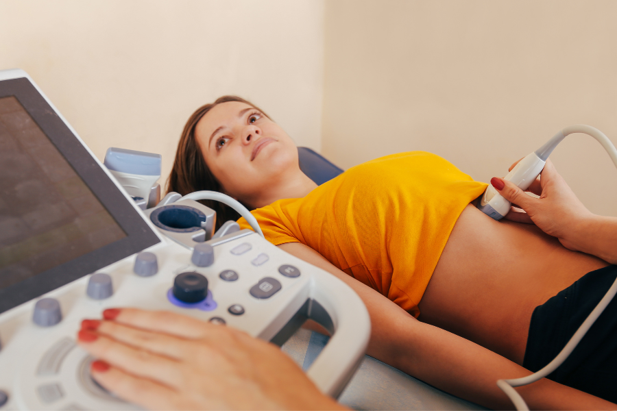
During the ultrasound, you will lie on a table. The vascular technologist will then apply a warm gel on your abdomen. The gel helps the ultrasound capture a clear picture, allowing the transducer wand to roll smoothly across your abdomen. As the technician gently sweeps the wand over your body, the machine will capture images and send them to a computer screen.
You may feel pressure during the test, but it shouldn’t be painful unless your technician is pressing on a sore or tender area. If you experience any discomfort, let your technician know so they can adjust the wand to make you more comfortable.
Is an Abdominal Ultrasound Better Than an X-ray?
 An abdominal ultrasound and an abdominal X-ray are different tests that provide different information for your doctor.
Dr. Levitt says, “With ultrasound, a lot of times we can do a non-invasive, non-radioactive technique to find out what your problem is.”
For many patients, a key benefit of ultrasounds is that they do not have a lasting impact on your body or health, but still allow doctors to make accurate diagnoses. The fact that the procedure does not require radiation to look inside the body makes it safe for pregnant women or patients who require repeated testing.
By contrast, an abdominal X-ray uses low-dose radiation to create images of the bones and tissues within the abdomen. It’s a useful test for certain conditions, such as abdominal injuries or bowel obstructions. However, X-rays are not as effective as ultrasounds for detecting soft tissue abnormalities—additionally, they expose the patient to radiation, which can be harmful in high doses.
Abdominal ultrasounds are just one of the many advanced diagnostic tools used by the clinical team at Vascular Specialists of Central Florida.
Backed by years of experience, our experts use ultrasound imaging to pinpoint underlying vascular conditions, mapping different areas of the body to ensure you stay happy and healthy.
Have questions about one of our procedures?
Don’t hesitate to call our office at 407-648-4323.